

A baby’s heart begins to form just a few weeks after conception. The cardiac tissue typically starts to pulse and beat around the 5th or 6th week of pregnancy. During this early stage, the heartbeat is often detectable using an early transvaginal ultrasound. By the 10th week, the fetal heart is fully developed into four chambers, and its rhythm can usually be heard clearly on a standard Doppler ultrasound.
A baby’s heart begins to form just a few weeks after conception. The cardiac tissue typically starts to pulse and beat around the 5th or 6th week of pregnancy. During this early stage, the heartbeat is often detectable using an early transvaginal ultrasound. By the 10th week, the fetal heart is fully developed into four chambers, and its rhythm can usually be heard clearly on a standard Doppler ultrasound.
The Emotional Gravity of the First Heartbeat
Hearing the rapid, rhythmic swoosh of your baby’s heartbeat for the first time is a profoundly transformative milestone. Before that moment in the ultrasound room, pregnancy can often feel like an abstract concept—a collection of physical symptoms, rising hormone levels, and changing dietary needs. But the sudden sound of that tiny, galloping rhythm instantly turns the abstract into a physical reality. It offers immense reassurance to expectant parents that a new life is actively growing and thriving within.
Understanding exactly how and when this vital organ develops provides incredible insight into the rapid pace of embryonic growth. The timeline of cardiogenesis (the formation of the heart) is a biological marvel. Within a matter of weeks, microscopic cells self-organize into a complex, pumping muscle that will sustain your child for a lifetime. This comprehensive guide breaks down the precise week-by-week development of the fetal heart, the cutting-edge medical tools used to detect it, and what you can expect during every major stage of your pregnancy journey.
The Biological Blueprint: How the Fetal Heart Forms
To truly appreciate when a baby’s heartbeat starts, you must look at the biological hardware being constructed right after fertilization. The cardiovascular system is the very first organ system to function in a human embryo. Because the rapidly growing cluster of cells quickly becomes too large to survive on simple nutrient diffusion alone, it desperately needs a dedicated delivery system for oxygen and nutrients.
The Formation of the Heart Tube
Around the third week of pregnancy (just days after a missed period), the embryo consists of three distinct layers of cells: the ectoderm, the mesoderm, and the endoderm. The heart and the primitive circulatory system begin to form within the middle layer, the mesoderm.
Specialized cells in the cardiogenic area begin to fuse, forming two parallel tubes. By the beginning of the fourth week, these two tubes merge into a single, primitive heart tube. Even at this incredibly early stage, before the structure resembles anything like a human heart, this primitive tube begins to contract.
The Crucial “S” Loop
The magic truly begins around the fifth and sixth weeks of gestation. The primitive heart tube begins to grow so rapidly that it runs out of space in the developing chest cavity. To accommodate its own explosive growth, the tube folds in on itself, bending into a distinct “S” shape. This complex folding process is critical; it creates the structural foundation for what will soon become the right and left sides of the heart. If the tube fails to loop correctly during this narrow window of development, it can result in congenital heart defects.
Month-by-Month Fetal Heart Development Timeline
The progression from a simple, pulsing tube to a fully functional, four-chambered heart happens at breakneck speed during the first trimester. Here is the week-by-week breakdown of your baby’s cardiovascular milestones.
Week 5: The Initial Pulses
By the fifth week of pregnancy, the fused heart tube begins its first spontaneous contractions. These early “heartbeats” are technically the result of electrical depolarization triggering the primitive muscle tissue to pulse. At this stage, the heart is pumping a microscopic amount of blood throughout the embryo. If you undergo a transvaginal ultrasound at five weeks, a highly trained technician might be able to spot the faint visual flicker of these contractions on the monitor, though it is usually too early to hear the sound.
Week 6: Detectable Rhythms
The sixth week marks a major turning point. The heart tube has successfully folded into its “S” shape, and the cardiac tissue is contracting at a much more regular, rapid rhythm—usually between 90 and 110 beats per minute. This is the stage where the heartbeat becomes significantly easier to detect visually on a transvaginal ultrasound screen.
Weeks 7 to 8: Chambers and Valves Emerge
During the seventh and eighth weeks, the internal architecture of the heart takes shape. The single tube begins to separate into four distinct chambers: the two upper receiving chambers (the atria) and the two lower pumping chambers (the ventricles). Simultaneously, the delicate tissue flaps that will become the heart valves begin to grow between these chambers, ensuring that blood flows in only one direction. The heart rate accelerates dramatically during these weeks, often peaking around 140 to 170 beats per minute.
Weeks 9 to 10: A Fully Formed Structure
By the end of the tenth week of pregnancy, the embryo officially graduates to fetal status. The fetal heart is now fully formed. It features four complete chambers, functioning valves, and major blood vessels connecting it to the rest of the body. While the heart will continue to grow in size and strength over the next 30 weeks, the intricate structural engineering is entirely complete. At this point, the heartbeat can typically be heard clearly using a handheld Doppler device in your obstetrician’s office.
Fetal Circulation: A Temporary Masterpiece
The heart beating inside your womb operates very differently than the heart beating inside your chest. Because a fetus is suspended in amniotic fluid, they do not use their lungs to breathe air. Instead, they receive entirely oxygenated blood directly from the mother via the umbilical cord and placenta. To accommodate this underwater environment, the fetal heart utilizes temporary anatomical shortcuts called shunts.
The Foramen Ovale
The first major shortcut is the foramen ovale, a temporary opening in the wall (the septum) between the right and left upper chambers of the heart. When oxygen-rich blood enters the right side of the fetal heart from the umbilical cord, the foramen ovale allows the blood to bypass the right ventricle and flow straight into the left side of the heart, where it is immediately pumped to the developing brain and body.
The Ductus Arteriosus
The second shortcut is a specialized blood vessel called the ductus arteriosus. Any blood that does happen to enter the right ventricle is pumped toward the lungs. However, since the lungs are collapsed and not yet functioning, the ductus arteriosus diverts this blood away from the pulmonary artery and pushes it directly into the aorta.
The moment a baby is born and takes their first massive gasp of room air, the pressure in their lungs drops. This pressure shift causes the foramen ovale to snap shut and signals the ductus arteriosus to permanently close over the next few days. The baby’s circulation instantly rewires itself to function just like an adult’s.
How and When Doctors Detect the Heartbeat
Medical technology has advanced to the point where healthcare providers can monitor fetal cardiovascular health with astonishing precision. Depending on how far along you are in your pregnancy, your doctor will utilize different tools to evaluate the heartbeat.
Transvaginal Ultrasounds (Early Pregnancy)
In the very early stages of pregnancy (between 5 and 8 weeks), the uterus is still tucked deep within the mother’s pelvis, behind the pelvic bone. A standard abdominal ultrasound cannot penetrate through the bone and dense tissue to see a microscopic embryo. Instead, doctors use a transvaginal ultrasound, which is inserted directly into the birth canal. This places the sound wave transducer just millimeters away from the developing uterus, allowing the technician to clearly see the visual fluttering of the 5-week-old cardiac tube.
Fetal Doppler (End of the First Trimester)
Right around the 10-to-12-week mark, the growing uterus pops up and out of the pelvic cavity. At this stage, your doctor will begin using a handheld fetal Doppler during your routine prenatal visits. They will apply a cool conductive gel to your lower abdomen and glide the wand across your skin. The Doppler uses sound waves to detect the movement of the baby’s pumping heart and translates that movement into an audible sound you can hear amplified through a speaker.
Fetal Echocardiogram (Second Trimester)
If your doctor suspects any abnormalities, or if you have a family history of congenital heart defects, they may order a fetal echocardiogram between 18 and 24 weeks of pregnancy. This is a highly specialized, in-depth ultrasound focused entirely on the baby’s heart. A pediatric cardiologist will analyze the blood flow, the integrity of the chamber walls, and the function of the valves to ensure everything is developing perfectly.
Normal Fetal Heart Rates By Week
A healthy fetal heart rate is not static; it fluctuates wildly depending on the stage of development and the baby’s activity level in the womb.
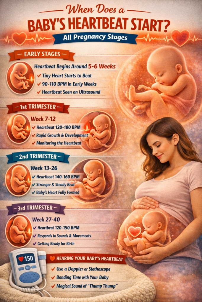
- 6 Weeks Gestation: The heart begins beating at a relatively moderate pace of 90 to 110 beats per minute (bpm).
- 9 to 10 Weeks Gestation: As the central nervous system rapidly develops, the heart rate skyrockets, often peaking between 140 and 170 bpm. It will sound incredibly fast, almost like a galloping horse.
- Second and Third Trimesters: As the heart muscle matures and the nervous system gains better regulation, the baseline heart rate settles into a steady average of 110 to 160 bpm.
Just like an adult’s heart rate spikes during a jog, a fetal heart rate will temporarily accelerate when the baby is actively rolling, kicking, or stretching in the womb.
What If You Cannot Hear the Heartbeat?
There are few things more terrifying than lying on an examination table while your doctor silently searches for a heartbeat. However, if no heartbeat is immediately detected early in your pregnancy, it is absolutely vital to avoid panicking. There are several highly common, non-threatening reasons why a heartbeat might be hiding.
Incorrect Gestational Dates
The most frequent reason a heartbeat cannot be found is simply that your dates are slightly off. Ovulation is not an exact science. If you ovulated just a week later than you thought, you might believe you are 7 weeks pregnant when you are actually only 6 weeks pregnant. During the first trimester, a delay of just five days makes a massive difference in what is visible on an ultrasound screen.
Tilted Uterus
A retroverted, or tilted, uterus tips backward toward the spine rather than forward toward the abdomen. This anatomical variation is completely normal and affects nearly 20% of women. However, early in pregnancy, a tilted uterus positions the tiny embryo further away from the ultrasound wand, making the faint heartbeat incredibly difficult to pick up until the uterus grows larger and pops forward later in the first trimester.
Maternal Body Composition
The ultrasound sound waves must travel through maternal skin, fat, and muscle to reach the uterus. A thicker abdominal wall can significantly muffle the sound waves of a handheld Doppler, often delaying the ability to hear the heartbeat until the 14th or 16th week of pregnancy.
The Position of the Placenta
If your placenta attaches to the front wall of your uterus (an anterior placenta), it acts as a massive, thick soundproofing cushion between the baby’s heart and the Doppler wand. The rushing sound of your own blood flowing through the placenta can easily drown out the faint, rapid ticking of the fetal heart.
Comparison Guide: Heartbeat Detection Methods
Use this quick-reference table to understand how and when medical professionals monitor your baby’s cardiac development.
| Detection Method | Earliest Week Used | How It Works | Best For |
|---|---|---|---|
| Transvaginal Ultrasound | 5 to 6 Weeks | Wand inserted into the birth canal for close-proximity imaging. | Confirming early viability, spotting the very first cardiac pulses visually. |
| Transabdominal Ultrasound | 8 to 10 Weeks | Wand glided across the lower stomach using conductive gel. | Routine anatomy checks, measuring crown-rump length, visual heartbeat confirmation. |
| Handheld Fetal Doppler | 10 to 12 Weeks | Bounces sound waves off the moving heart valves to produce audio. | Routine, quick in-office checks to listen to the heartbeat sound. |
| Fetal Echocardiogram | 18 to 24 Weeks | Deep, targeted 3D/4D ultrasound isolating the cardiac anatomy. | Diagnosing structural heart defects, checking blood flow and valve function. |
| Fetoscope | 20+ Weeks | A specialized stethoscope pressed against the maternal abdomen. | Low-tech, non-electronic heartbeat detection by midwives late in pregnancy. |
Tracking the Broader Pregnancy Stages
While the heartbeat is the most anticipated milestone, it is just one small piece of the monumental biological marathon taking place inside your body. Pregnancy is divided into three trimesters, each characterized by distinct developmental leaps.
The First Trimester: Organogenesis (Weeks 1-13)
The first trimester is the era of construction. Known medically as organogenesis, this is the period when every major organ system is rapidly built from scratch. Alongside the beating heart, the neural tube closes to form the brain and spinal cord. Tiny arm and leg buds erupt and elongate into limbs with webbed fingers and toes. The face begins to take shape, forming depressions for the eyes and nostrils. Because this foundational construction is so complex, the first trimester is the most sensitive period for the developing embryo, making early prenatal care and avoiding harmful substances critical.
The Second Trimester: Rapid Growth and Quickening (Weeks 14-27)
Often called the “golden trimester,” this phase is characterized by intense growth and maturation. The organs are completely formed and now simply need to grow in size and practice their functions. The kidneys begin producing urine, and the bone marrow starts producing red blood cells. Most excitingly, the baby’s skeleton hardens from soft cartilage into bone, allowing their movements to become forceful enough to feel. This sensation, known as quickening, usually occurs between 16 and 20 weeks. By the halfway point (week 20), you will typically undergo a comprehensive anatomy scan to measure the baby’s growth and confirm the sex.
The Third Trimester: Maturation and Fat Accumulation (Weeks 28-40)
The final stretch is all about gaining weight and preparing for the outside world. The baby rapidly packs on adipose (fat) tissue, which will help them regulate their body temperature after birth. The brain grows exponentially, developing deep grooves and folds. The most critical milestone of the third trimester is lung maturation. The lungs begin secreting surfactant, a slippery substance that keeps the air sacs open so the baby can breathe oxygen. As the baby runs out of room in the expanding uterus, the wild somersaults transition into deep, rolling movements and sharp jabs.
Preparing for Your Baby’s Arrival
As your pregnancy progresses and you move past the anxiety of the early ultrasounds, you can finally shift your focus toward welcoming your child home. Nesting is a powerful biological urge, and channeling that energy into preparing your environment is highly productive.
You do not have to navigate the transition into parenthood alone. Connecting with other expectant parents and relying on trusted advice from the Wobblebee parenting community provides invaluable support as you journey toward your delivery date.
Frequently Asked Questions
Can I hear my baby’s heartbeat with a regular stethoscope? It is incredibly difficult to hear a fetal heartbeat with a standard medical stethoscope until very late in the third trimester. Before 20 weeks, the baby is simply too small, and the thick layers of amniotic fluid, uterine muscle, and maternal abdominal tissue muffle the delicate sound entirely.
Why is my baby’s heart rate so fast during an ultrasound? A fetal heart beats much faster than an adult heart, normally ranging between 110 and 160 beats per minute. This rapid rate is a perfectly healthy sign of a maturing nervous system, and the speed will naturally peak around the 9th or 10th week before slowly stabilizing in the second trimester.
Can the fetal heart rate predict my baby’s gender? No. Despite popular old wives’ tales claiming that a heart rate above 140 bpm means you are having a girl and below 140 bpm indicates a boy, extensive medical studies have proven there is zero correlation between fetal heart rate and the biological sex of the baby.
Is it safe to use an at-home fetal Doppler? Most medical professionals strongly advise against using at-home, over-the-counter fetal Dopplers. Without proper medical training, parents frequently mistake the rushing sound of the placenta or their own maternal pulse for the baby’s heartbeat, leading to unnecessary panic or a false sense of security.
What happens if no heartbeat is found at an 8-week ultrasound? If a transvaginal ultrasound definitively confirms a gestational sac and a fetal pole measuring over 7 millimeters but detects no heartbeat, it sadly indicates an early pregnancy loss. However, your doctor will almost always schedule a follow-up scan a week later to absolutely confirm the diagnosis before discussing your medical options.